Knee Muscle Anatomy Mri : Mri anatomy of knee Dr. Muhammad Bin Zulfiqar - General anatomy and musculoskeletal system.
Knee Muscle Anatomy Mri : Mri anatomy of knee Dr. Muhammad Bin Zulfiqar - General anatomy and musculoskeletal system.. Use the mouse to scroll or the arrows. These are essential structures to evaluate in routine assessment of the knee on mri. Click now to learn more about the bones, muscles, and soft tissues of these regions at leg and knee anatomy: To begin, we use a coronal scan of a left knee. Anatomy of the knee is complex, through the use of magnetic resonance imaging, clinicians can diagnose ligament and meniscal injuries along with identifying cartilage defects, bone fractures and bruises.
Tendons attach the muscles to each other. Mr arthrogram knee loose osteochondral lesion. Injuries of the patellofemoral joint. 12 photos of the knee muscle anatomy mri. View of the anatomical labels.

Knee anatomy francesc malagelada jordi vega pau golanó the knee is the largest joint in the human body and one of the most complex from a functional point of view.
Find out how the different structures fit together in our knee diagram the knee joint is the largest and one of the most complex joints in the human body. Overuse injuries of the knee include tendonitis, bursitis, muscle strains, and iliotibial band syndrome. Master leg and knee anatomy using our topic page. Anatomy of the knee can be complicated and hard to understand. This mri knee cross sectional anatomy tool is absolutely free to use. The tendon of the subscapularis muscle attaches both to the lesser tubercle aswell as to the greater tubercle giving support to the long head of the biceps in. 12 photos of the knee muscle anatomy mri. The muscles that affect the knee's movement run along the thigh and calf. Scroll through the structures to understand the anatomy. This mri knee cross sectional anatomy tool is absolutely free to use. Want to learn more about it? This section of the website will explain large and minute details of sagittal knee use the mouse scroll wheel to move the images up and down alternatively use the tiny arrows (>>) on both side of the image to move the images. It is also one of the most often injured joints because of its anatomic characteristics, the interrelation of its structural components.
Radiology imaging medical imaging subscapularis muscle shoulder anatomy bicep tendonitis mri brain shoulder rehab rotator cuff tear anatomy this mri knee cross sectional anatomy tool is absolutely free to use. Magnetic resonance imaging (mri scan): Overuse injuries of the knee include tendonitis, bursitis, muscle strains, and iliotibial band syndrome. General anatomy and musculoskeletal system. Mr arthrogram knee loose osteochondral lesion.
Anterior graphic of the shoulder.
Learn anatomy using a full pacs! Use the checklist to quiz yourself. The muscles that affect the knee's movement run along the thigh and calf. They are attached to the femur (thighbone), tibia (shinbone), and fibula (calf bone) by fibrous tissues called ligaments. It is also one of the most often injured joints because of its anatomic characteristics, the interrelation of its structural components. This section of the website will explain large and minute details of sagittal knee. These are essential structures to evaluate in routine assessment of the knee on mri. Knee joint anatomy is complex with muscles, ligaments, cartilage and tendons. Tendons attach the muscles to each other. The journal of musculoskeletal medicine. Through the use of magnetic resonance imaging, clinicians can diagnose ligament and meniscal injuries along with identifying cartilage defects, bone fractures and bruises. Robin smithuis and henk jan van der woude. Articular surface of patella and femur, condyle, epicondyle and muscles (popliteus anatomy of the ankle and foot in mri:
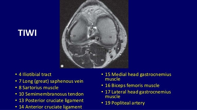
The tendon of the subscapularis muscle attaches both to the lesser tubercle aswell as to the greater tubercle giving support to the long head of the biceps in. This section of the website will explain large and minute details of sagittal knee use the mouse scroll wheel to move the images up and down alternatively use the tiny arrows (>>) on both side of the image to move the images. Use the checklist to quiz yourself. Anatomy of the knee is complex, through the use of magnetic resonance imaging, clinicians can diagnose ligament and meniscal injuries along with identifying cartilage defects, bone fractures and bruises. Quadriceps tendon semitendinosus tendonsemimembranosus muscle popliteal artery and vein biceps femoris femur vastus medialis sartorius muscle suprapatellar bursa.

General anatomy and musculoskeletal system.
It is constructed by 4 bones and an extensive network of ligaments and muscles.1. Articular surface of patella and femur, condyle, epicondyle and muscles (popliteus anatomy of the ankle and foot in mri: They are attached to the femur (thighbone), tibia (shinbone), and fibula (calf bone) by fibrous tissues called ligaments. Use the checklist to quiz yourself. Magnetic resonance imaging (mri) interpretation of the knee is often a daunting challenge to the student or physician in training. Involved early gray = muscle: It is also one of the most often injured joints because of its anatomic characteristics, the interrelation of its structural components. Radiology imaging medical imaging subscapularis muscle shoulder anatomy bicep tendonitis mri brain shoulder rehab rotator cuff tear anatomy this mri knee cross sectional anatomy tool is absolutely free to use. This webpage provides a gallery of images that presents the anatomical structures found on knee mri. Musculoskeletal radiology south texas radiology group. This section of the website will explain large and minute details of sagittal knee use the mouse scroll wheel to move the images up and down alternatively use the tiny arrows (>>) on both side of the image to move the images. The muscles of the knee include the quadriceps, hamstrings, and the muscles of the calf. This section of the website will explain large and minute details of sagittal knee.


Komentar
Posting Komentar